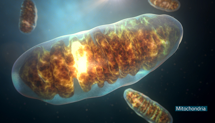
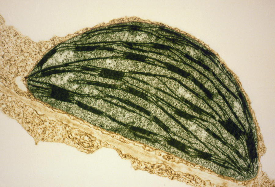
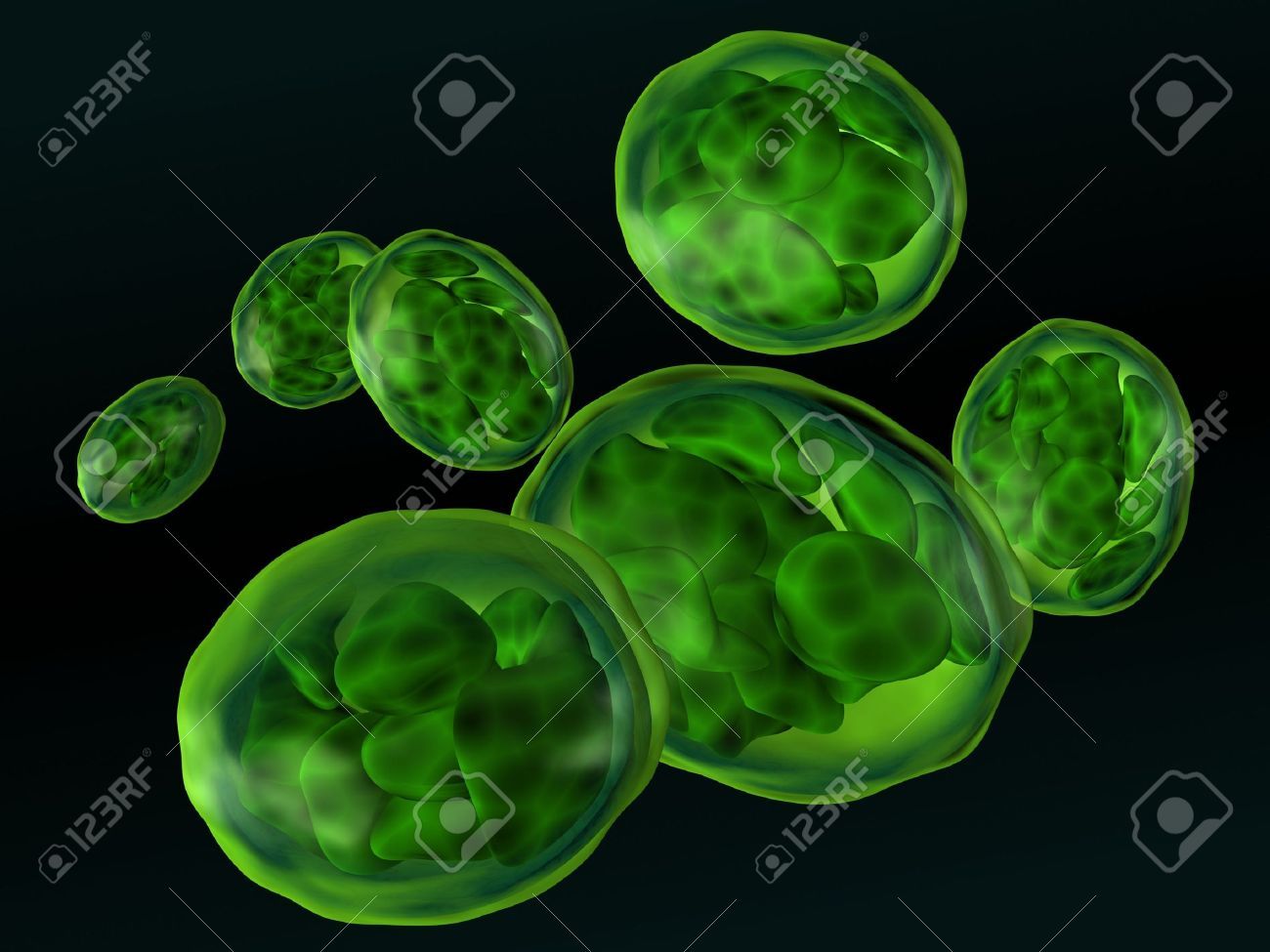
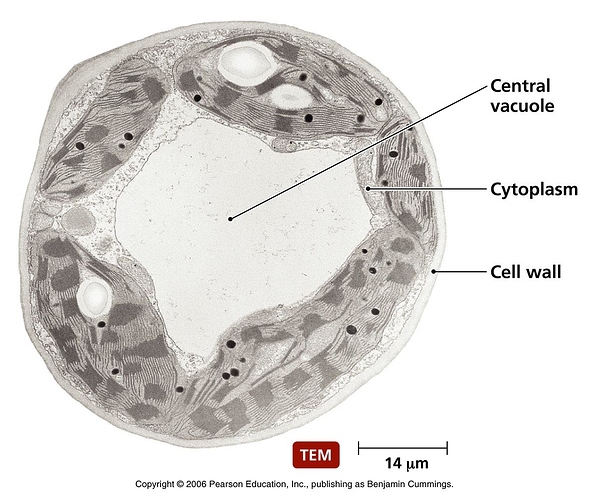
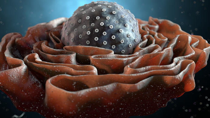
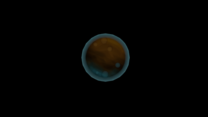
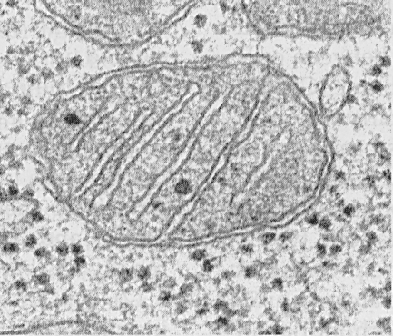
I am still on vacation, so sorry if I take too long to reply to any posts. For inspiration to the modelers, I have compiled a list of the necessary organelles with their actual real-life photographs and a cool looking stock image.
Mitochondrion
Chloroplast
We currently have one from Chizu: http://i.imgur.com/ZClpC7Ib.jpg but it is untested (although I really like it)
Vacuole
As you can see it is more-or-less transparent and can be of any shape, not necessarily round. We already have one from tjblazer85: https://cloud.githubusercontent.com/assets/4619968/6841437/8fa63e48-d3c5-11e4-8505-b355da82e6b3.png but the material was not working correctly, so if anyone wants to fix it or make their own, go for it. We also need a lot of different vacuoles for all the different hex arrangements (AKA 2 hexes, three hexes forming a triangle, three hexes in a line, etc.)
Nucleus with endoplasmic reticulum
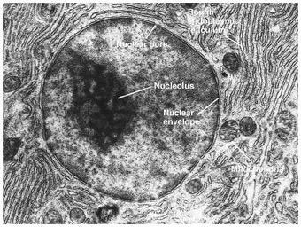
and the Golgi Apparatus
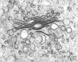
We need the nucleus, ER, and Golgi Complex to be one mesh that spawns 6 hexes (3 by 2). As you can see, the endoplasmic reticulum looks identical to the ER, and it is! They are made of the exact same material and have the exact same structure. As such, it would be impossible to tell them apart if they were all combined and coloring them different would just be unrealistic. I suggest that the person who decides to model this either makes 2 different meshes—Nucleus+ER in one and GA in the other—or simply makes one mesh with a gap between the Nucleus+ER and GA. Also, note that the half of the Endoplasmic Reticulum closer to the nucleus should have dots on it that symbolize ribosomes while the other half should be smooth and have “membrane bubbles” that are barely attached, if at all, to the ER.
Here is a render:
I assume we already have models for Flagella and Cilia so no need to make ones for those. The Agent Vacuole should look similar to the normal vacuole, but be of a different color. I also believe it should be attached to the cell membrane, but this is up for discussion. Here is the one from Tritium that we have so far:
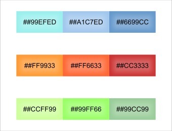
As you can obviously see, the SEM photographs are all black-and-white, so you have a lot of artistic freedom when deciding how they should look. Here is a table of colors we are currently using in Thrive that you could use as a reference:
Finally, since we are on the topic of cells and their models, here is a video from http://joelotron.com/:
https://vimeo.com/123332533
This guy makes some seriously awesome cell and bacteria renders.
